- One in every four to six women in Europe affected by fibroids
- Ultrasound enables diagnosis and opens the door to treatments that preserve fertility, allowing women to realise their desire to have children.
- Live scans and patient feedback – top-level educational events at the ISUOG World Congress

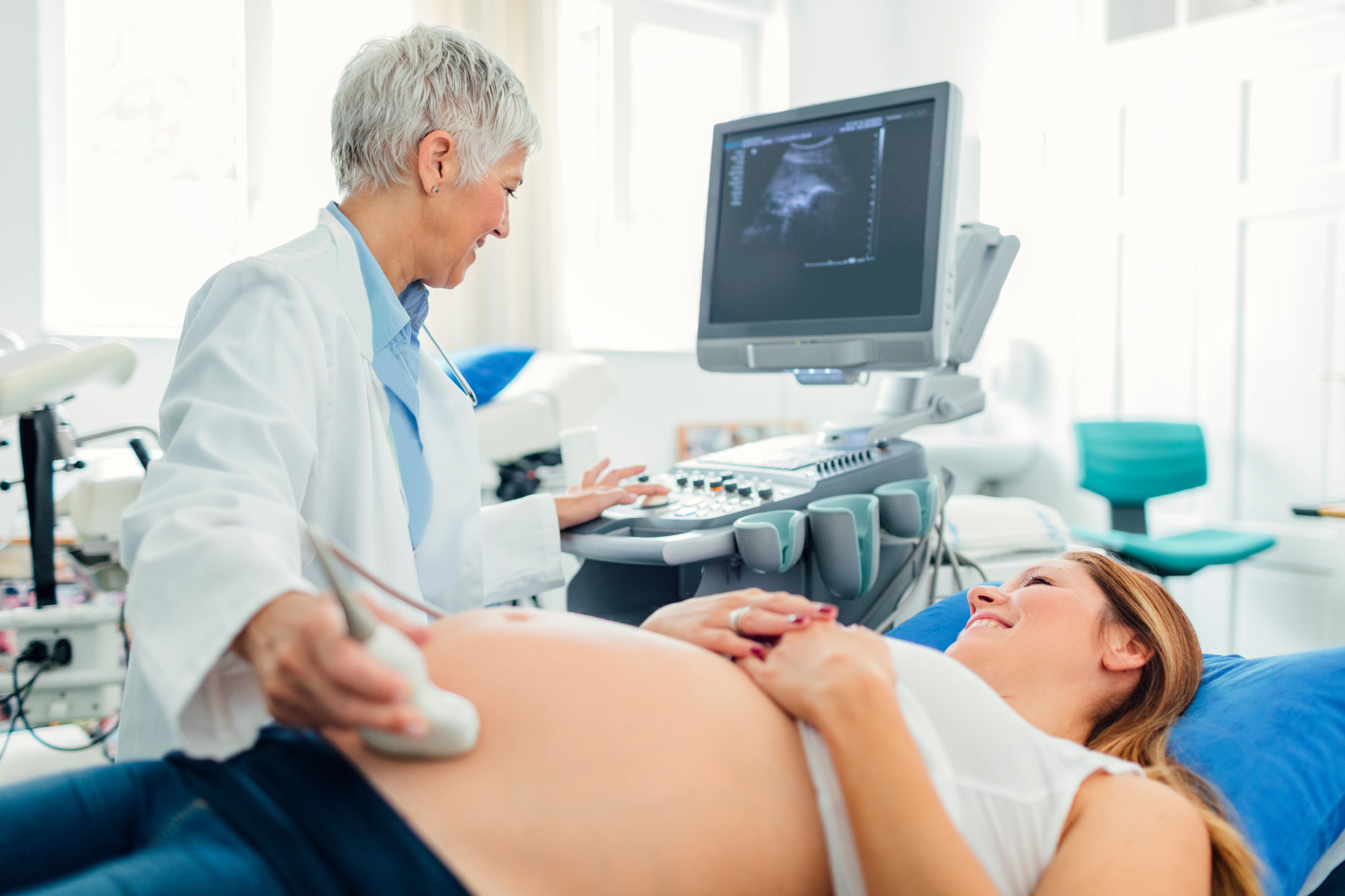
“Ultrasound examinations are an indispensable part of modern-day obstetrics and gynaecology. As well as being used in regular check-ups to monitor the development of the embryo during pregnancy, ultrasound is playing an increasingly important role in treating young women with endometriosis and fibroids. The insights delivered by this imaging technique make it possible to respond earlier, preserve women’s fertility and make future pregnancy possible,” explained Dr. Christoph Brezinka from the University Hospital for Gynaecological Endocrinology and Reproductive Medicine at the Medical University of Innsbruck.
Ultrasound enables identification, monitoring and treatment of fibroids
Around one in every four to six women of child-bearing age sufferers from fibroids. These growths that develop in the wall of the uterus are among the most common type of benign tumour found in the female reproductive system. While in the 1980s removal of the womb was the standard procedure following diagnosis, developments in modern ultrasound technology now mean that it is possible to monitor and treat patients with medication first. If surgery is required, the uterus can be kept, preserving fertility. “Of course, in some cases targeted treatments are required to support the desire to have children,” explained Brezinka. “But the fact that the young women affected by the condition are still in a position to conceive is a huge step forward.”
Ultrasound used to diagnose and monitor the progression of endometriosis
Similar advances have also been made in the treatment of endometriosis – a chronic, benign condition also found in reproductive-age women in which the tissue that makes up the lining of the womb is present on other organs inside the body, causing symptoms such as irregular bleeding, cysts and painful inflammation. 10-15% of all women of childbearing age are affected by the condition. While diagnosis was previously only possible following invasive surgery, nowadays ultrasound can be used to diagnose and monitor the progression of the disease. Therapies are adapted depending on whether the patient wishes to have children. “Diagnosis based on ultrasound examinations is critical for health of women and their ability to have children in future,” Brezinka explained.
Live scans and patient sessions – top-level educational events
As doctors have very little time in their day-to-day work to keep on top of all the latest developments in ultrasound technology, participating in the ISUOG World Congress makes a great deal of sense. To provide practical training, various experts will demonstrate the full functionality of the latest generation of ultrasound equipment in live sessions. Self-help groups for uterine fibroid, endometriosis and tumour patients are also invited to participate in discussions, with a view to bringing about specific improvements in ultrasound examinations for women with these conditions.
Around 2,000 prenatal diagnosticians and specialists will meet at the Austria Center Vienna between 15 and 21 September 2017 for the International Society of Ultrasound in Obstetrics and Gynaecology (ISUOG) World Congress to discuss the latest research findings in their discipline. The Austrian congress presidents are Dr. Christoph Brezinka from the University Hospital for Gynaecological Endocrinology and Reproductive Medicine in Innsbruck and Dr. Daniela Prayer, Head of the Division of Neuroradiology and Musculoskeletal Radiology at Vienna General Hospital.


